Tuesday 15 December 2015

A Joyous, Healthy and Active Festive Season

Once again I would like to thank all my loyal readers for your continued support throughout the year. I hope you have gained some useful insight from the topics covered during the past year.

I wish you a joyous festive season and a prosperous new year. May 2016 be a happy and healthy year for you all.

Remember to keep active during the festive season! Play and laughter can be the best exercise! J

Monday 2 November 2015

Plantar Fasciitis

Plantar fasciitis is one of the most common foot injuries, affecting approximately 10% of runners. It is an inflammatory condition, in which the connective tissue on the underside of the foot becomes inflamed. Intrinsic factors contributing to this condition include biomechanical factors related to the foot and ankles, as well as muscle imbalances in strength and flexibility of the lower leg and foot muscles. Extrinsic factors include incorrect footwear, poor training and excessive running on hard or uneven surfaces.

Signs and Symptoms
Pain is experienced on the underside of the foot, near the heel, and is usually worse after rest and will be particularly severe first thing in the morning when weight is placed on the foot. This pain tends to improve within a few minutes of walking. In more severe cases, where there is neural involvement as well, pain is more severe and widespread. Pain often improves with activity, but will recur after rest.

Management
Ice and non-steroidal anti-inflammatory drugs are used to manage the acute pain and inflammation. Massage is used to reduce inflammation in the fascia. Strapping and heel lifts may be used to take the pressure off the arch of the foot. Achilles tendon stretches should be performed, as well as various other exercises to improve range of motion in the ankle and foot, within pain-free ranges. Gentle strengthening exercises can be done to improve muscle strength around the foot and ankle, again within pain-free ranges. A biokineticist can then assess factors contributing to the condition and provide appropriate stretching and strengthening exercises to improve muscle imbalances, thereby preventing the condition form recurring.

References
Foundations of Athletic Training: Prevention, Assessment and Management

Tuesday 13 October 2015

Newsflash!!

I would like to welcome Monique Turner to my practice.
Monique is a registered biokineticist and she will be practicing at:
95 Boeing Road East
Bedfordview
(entrance on Marais Street)
Tel: 011 454 0232

Tuesday 6 October 2015

Hamstring Strains

The hamstrings are the most commonly strained muscles in the body. This injury occurs when the hamstrings are stretched beyond their acceptable range of movement, either in a sudden movement, such as a violent stretch, or from repetitive motions. Hamstring strains range from mild (Grade I) to severe (Grade III), depending on the number of muscle fibres affected and the severity of the strain.

Risk factors for hamstring strains
The following factors increase the risk of developing a hamstring strain:
·         Poor flexibility
·         Poor posture
·         Muscle strength imbalance
·         Improper warm-up
·         Muscle fatigue
·         Poor neuromuscular control
·         Previous injury
·         Overuse
·         Incorrect technique

Signs and symptoms
Grade I (mild) hamstring strain:
·         Mild pain and tightness in the back of the thigh
Grade II (moderate) hamstring strain:
·         Tearing or popping sensation
·         Moderate pain and weakness
·         Difficulty bending the knee
Grade III (severe) hamstring strain:
·         Tearing or popping sensation
·         Severe pain and weakness
·         Individual will limp
·         Inability to straighten the knee completely or strike the ground with the heel first when walking
·         Difficulty bending the knee
·         Swelling and bruising occurs 1-2 days after the injury

Management
Initial treatment should include rest, ice, compression and elevation. Non-steroidal anti-inflammatory drugs are often prescribed as well. Crutches are used in more severe cases, if deemed to be necessary. Physiotherapy is required to reduce pain and swelling and regain range of movement.
Individuals with this injury often complain that the injury is chronic and recurring. This is usually because the strengthening and final-phase rehabilitation is neglected. It is crucial that the hamstrings are strengthened following a strain, to prevent the injury from reoccurring and to prevent injury to other parts of the body, such as the knee, as a result of muscle imbalances. A biokineticist can assist in prescribing appropriate exercises to strengthen the hamstrings and return an individual to full function, whether it be the normal daily activities, or specific sporting movements.

References
Foundations of Athletic Training: Prevention, Assessment and Management

Friday 4 September 2015

Ankle Joint Fusion

Fusion of the ankle joint may be required as a result of chronic degeneration of the joint from conditions such as osteoarthritis. A surgical procedure is performed, where the bones are fused together, reducing movement and, therefore, pain at the joint. This loss of mobility of the ankle joint can have numerous side-effects on balance, walking pattern and functional ability. Recovery from an ankle fusion is long and includes the ankle being immobilized and the patient being off the affected leg for several weeks, sometimes as long as 10-12 weeks. The patient is then put into a moon boot and partial weight-bearing will be allowed for several more weeks. Once your surgeon is satisfied that the bones have completely fused, you will be able to return to more normal activities. This prolonged recovery has significant implications on the surrounding leg muscles, joints, balance and walking pattern.

If you have had an ankle fusion, it is important to consider the implications of the fusion on the rest of your body. The lack of mobility at the joint itself will affect how you walk and your ability to carry out your daily activities, such as climbing stairs. Your posture may also be affected. These changes may cause aches and pains in other areas of your body, because you are now walking and standing differently to what you are used to. Adaptations to one’s walking pattern and functional movements will most likely need to be made to compensate for the restricted movement at the ankle joint.

The muscles of the calves and thigh will have become atrophied, or wasted, due to lack of use while the affected leg was not being used so that it could heal. It is important to rebuild the strength of these muscles following an ankle fusion, so that walking and functional ability can return to as close to normal, as soon as the fused joint will allow. Strengthening of the leg and core muscles will assist in the adaptation process of getting used to the new joint.

A biokineticist will assess and provide appropriate exercises to assist in rebuilding the leg muscles, regaining balance, and re-establishing a natural and correct posture and walking pattern. If the appropriate rehabilitation programme is followed, one will be able to return to most previous activities, with only slight adaptations needing to be made.

References
http://www.aofas.org/footcaremd/treatments/Pages/Ankle-Arthrodesis.aspx

Thursday 6 August 2015

Lower-Limb Amputation

Lower-limb amputation refers to the surgical removal of the lower limb (leg) or part of the lower limb. Of course, amputations of the upper limb, or arm, also exist, but due to the non-weight-bearing nature of the arm, this is much less complicated and so will not be discussed in this blog article.

Lower-limb amputations are caused by the following:
·         Circulatory and vascular diseases associated with type 2 diabetes and peripheral vascular disease – 70%
·         Trauma to the limb itself – 23%
·         Removal of tumours – 4%
·         Congenital deformities – 3%

Lower-limb amputations caused by vascular diseases are more common in individuals over the age of 55 years; whereas, those caused by trauma, tumours or congenital deformities are more common in individuals under the age of 50 years.

It is important to know the cause of an amputation from an exercise perspective because the purpose of the exercise therapy will differ slightly. If an amputation was caused by a vascular disease, then it is important that the exercise programme focuses on reducing the progression of the associated vascular condition. On the other hand, the purpose of the exercise programme for those with amputations caused by trauma, tumours or congenital deformities is the same as that for able-bodies individuals, the aim being, to reduce the risk of diseases associated with a sedentary lifestyle, such as high cholesterol, high blood pressure, diabetes and obesity. Individuals who have had an amputation are at greater risk of developing these diseases, as they are more likely to be sedentary, purely because of their physical limitation. However, this should not be an excuse not to exercise, as there is a wide variety of exercises that can still be performed.

Depending on the level of amputation – at the foot, below the knee, above the knee or at the hip – various factors must be considered when planning an exercise programme. Appropriate exercise modalities need to be chosen according to the individual’s physical capabilities and current level of fitness. Swimming and arm ergometry are generally both safe and appropriate forms of exercise for most lower-limb amputees. Strengthening exercises are important and can usually be done with minor adaptations. It is generally more safe to use machines, rather than free weights, because an amputee’s balance is more likely to be compromised.

Walking is important to enable individuals to become as independent as possible; however, one cannot overdo it. Individuals with a prosthetic leg are susceptible to sores and infections, as a result of the prosthesis rubbing on the skin. This can cause further disability if the sores do not heal. These individuals also expend more energy when walking than able-bodied individuals and so cannot walk as far or for as long. Phantom pain, a common complaint of amputees, is pain experienced in the amputated limb. This pain ranges from mild to severe and can also hinder one’s exercise ability.

Although amputation is considered a disability, there is still a wide range of movement and functional exercises that amputees can achieve, ranging from the normal daily activities to competitive sport. It is crucial that one does not give up following an amputation, but rather seeks professional advice, such as that of a biokineticist, to assist in implementing an appropriate exercise programme that focuses on the needs and goals of the individual.

References
ACSM’s Exercise Management for Persons with Chronic Diseases and Disabilities

Tuesday 7 July 2015

Hypermobility Syndrome

Many people experience hypermobility in one or more of their joints. This means that the joint can move beyond the expected normal ranges for that joint. This is often referred to as being “double-jointed”. Usually, this condition has no symptoms and one is able to lead a normal life, unless there is an injury to the joint, causing pain and/or inflammation. If numerous joints are involved and symptoms are present, then one is believed to have hypermobility syndrome. This syndrome has a strong genetic link and thus often runs in families.

Signs and Symptoms
As mentioned earlier, a hypermobile joint often has no symptoms, unless injury to that joint occurs. However, the symptoms of the syndrome include:
·         Pain in the knees, hips, elbows and fingers
·         Clicking in the joints
·         Recurring joint sprains and dislocations due to instability of the joints
·         Fatigue
·         Dizziness and fainting
·         Scoliosis is more common in individuals with hypermobility syndrome and this can result in back pain
·         Joint hypermobility decreases as we get older, as we become less flexible with age

The following signs can be seen in individuals with hypermobility syndrome:
·         The ability to hyperextend the knees and/or elbows past 10 degrees
·         The ability to stand with the palms flat on the floor while keeping the knees straight
·         The ability to touch the thumb to the forearm

Hypermobility syndrome is diagnosed by means of physical examination according to the above-mentioned signs, as well as various additional tests. There is no blood test or x-ray to diagnose this syndrome.

Treatment and Management
Because there are often no symptoms, treatment is often not required. The condition generally improves as individuals get older because flexibility of the joints decreases with age. If a joint has been sprained or dislocated, then that joint will be treated for that specific injury. The pain associated with hypermobility syndrome can be treated with appropriate pain management drugs according to your doctor. Exercise therapy is particularly important in managing this condition, as the muscles surrounding the joints can be strengthened to improve stability of the joints, thereby reducing the risk of injury. A biokineticist can assist in prescribing and monitoring an appropriate exercise programme that strengthens the muscles while minimizing the risk of risk of injury.

Prognosis
Generally, there are few or no long-term side effects of joint hypermobility. However, in severe cases, individuals may develop arthritis at a later stage. It is, therefore, important to manage the condition with appropriate strengthening exercises from an early age to prevent any injuries and long-term side-effects.

References

Thursday 4 June 2015

Cerebral Palsy

Cerebral Palsy (CP) is a non-progressive condition that results from a lesion in the brain that occurs either before, during, or shortly after birth, resulting in the abnormal development of the brain. Failure of the brain to develop properly can occur during the first or second trimesters of pregnancy and can be caused by genetic disorders or limited blood supply. A lesion may also develop as a result of injury to the brain before, during, or after birth, such as a traumatic blow to the head or bleeding in the brain.

Depending on the severity of the lesion and the part of the brain affected, individuals with CP will present with varying degrees of disability. However, the general characteristic features of CP are limited mobility, poor coordination, and a limited ability to maintain posture and balance. These individuals may be wheelchair bound, unable to walk at all, or may be able to walk, but with an abnormal gait, or walking pattern. They will also often present with spasticity in the hands and arms, and a misshapen mouth, resulting in drooling and slurred speech. Their cognitive function is usually not affected, therefore, it is important not to treat them as cognitively impaired simply because they drool and have a slurred speech.

Because of the varying degrees of disability in CP patients, every person must be assessed and treated as an individual according to his/her goals and degree of disability. Adaptations to the assessment and management of these individuals will need to be made to accommodate each individual’s needs and goals. Any medications that the individual is taking must be considered, as these may alter his/her response to exercise. It is important to realize that the lesion in the brain cannot be reversed or changed by exercise; however, exercise therapy can help to improve the symptoms of spasticity, poor balance and muscle weakness associated with CP. It is also important to note that a person with CP may experience increased spasticity and decreased coordination shortly after exercise, but this is not uncommon and will return to normal.

Individuals with CP are often sedentary, due to their lack of mobility. This increases their risk of developing sedentary diseases, such as cardiovascular disease and stroke. It is, thus, important that they embark on an appropriate, regular exercise programme in order to reap the physiological and psychological benefits of regular exercise. Improvements in mood and self-esteem are also associated with exercise, which the CP patient will benefit from. The exercise programme should incorporate all the major muscle groups (if still functional) and should be aimed at improving cardiovascular fitness, muscles strength and flexibility, thereby increasing one’s independence to carry out his/her daily activities and, in turn, improving one’s sense of self-worth. A biokineticist can assist in the design, implementation and adaptation of such an exercise programme.

References
ACSM’s Exercise Management for Persons with Chronic Diseases and Disabilities

Monday 4 May 2015

Anterior Cruciate Ligament (ACL) Rehabilitation

A common knee injury amongst the active population is the damage to or rupture of the anterior cruciate ligament (ACL). The role of this ligament is to stabilize the knee joint by stopping the bottom of the leg from collapsing forward under the knee joint, particularly during deceleration, landing, or turning and cutting actions. It is unusual for the ACL to be injured in isolation; often other ligaments and/or the meniscus are also injured. Those at greater risk of tearing the ACL are, therefore, those involved in jumping and pivoting sports, as well as contact sports, such as soccer and rugby. A muscle strength imbalance between the hamstrings and the quadriceps muscles (back and front of the thigh respectively) can also increase the risk of this injury occurring.

Signs and Symptoms
As the injury occurs, a popping, tearing, or snapping sensation may be heard or felt, followed by swelling around the knee joint. Pain may be felt immediately or occur later and may range from mild to severe. The pain may be felt deep in the knee joint or, more commonly, on the front side of the knee. Generally, one is able to walk on the affected leg; however, this is often associated with a feeling of the knee wanting to collapse or a general feeling of discomfort. Because of the high likelihood of other surrounding tissues being damaged as well, it is important to consult a specialist for further investigation.

Management
Depending on the severity of the damage to the ACL and the involvement of the surrounding structures, an ACL injury is either treated conservatively or surgically.

Conservative management involves rest, ice, compression, elevation and immobilization of the knee joint to reduce swelling. Crutches can be used if walking is too painful. Pain-free range of motion exercises are performed to maintain mobility in the joint. Gentle and appropriate strengthening exercise are done to maintain muscle strength. Physiotherapy is very important in these initial stages of recovery. Upper body exercises can be continued throughout the recovery period. Strengthening, range of motion and proprioception and balance exercises can be progressed appropriately by a biokineticist until the individual is back to full function. This can take anywhere between 6 weeks to 3 months, depending on the severity of the injury and the compliance of the individual with the rehabilitation programme.

If there is a complete rupture of the ACL or multiple surrounding tissues are also affected, surgery is usually recommended so that an individual can return to contact or pivoting-type sports. Depending on the other structures damaged, the repair and recovery will be slightly different. A graft is taken from the hamstring muscle to repair the ACL, which means that the hamstring muscle also needs to be carefully rehabilitated and strengthened to avoid a later hamstring injury. After surgery, the affected leg is immobilized in a full-leg brace, which can be locked at various degrees of flexion and extension. One will more than likely be non-weight-bearing for between 2 and 6 weeks, again depending on damage to other structures, such as the meniscus. Physiotherapy is crucial to maintain range of movement in the joint and reduce swelling. Again, gentle strengthening exercises are done to maintain muscular strength around the knee. Once the brace is removed, the rehabilitation will follow in a similar fashion to the conservative treatment plan, progressing appropriately with the guidance of a biokineticist, with an additional focus on the rehabilitation of the affected hamstring muscle. If properly rehabilitated, one should be able to return to sport if they have not lost the nerve to do so.

References
Foundations of Athletic Training: Prevention, Assessment and Management

Monday 30 March 2015

Polio and Post-Polio Syndrome

Poliomyelitis is an acute, viral, epidemic disease that attacks the motor neurons that control the leg muscles, resulting in paralysis and, therefore, atrophy of the affected muscles. The virus eventually clears itself and one recovers fully, leading a normal and active life. However, up to 40 years later, an individual who had contracted the virus previously can start to develop similar symptoms of fatigue, weakness, and muscle and joint pain that were experienced at the time of contraction. This is known as Post-Polio Syndrome (PPS) and it affects at least one quarter of those initially affected. Other possible symptoms include sleep disorders and intolerance to cold.

Depending on the severity of the virus, varying portions of the leg muscles will be affected, directly affecting one’s functional ability, strength and power. A limp is often present due to muscle loss in the leg, and balance is generally poor on the affected leg. The lack of use of the leg due to weakness further contributes to muscle wasting, thereby exacerbating symptoms.

Exercise is essential in the management of PPS; however, an appropriate and individualised programme is essential for success. The affected motor units may not increase in strength; however, the surrounding unaffected muscles can be strengthened to support and compensate for the affected muscles. Core and upper body strengthening exercises should be included to improve posture and gait (walking pattern). Appropriate whole body exercises should be done to include as many muscle groups as possible and increase physical fitness. Those with PPS are also more susceptible to depression and exercise plays an important role in managing depression, by increasing one’s sense of achievement and well-being. Fatigue is a common symptom amongst people with PPS, so an appropriate intensity with many rests must be prescribed. An individual with PPS should be able to continue with an appropriate exercise programme on a regular basis on his/her own, adjusting the programme accordingly. However, initially it is important to see an appropriate exercise specialist, such as a biokineticist, to be assessed and start an suitable exercise regime.

References
ACSM’s Exercise Management for Person’s with Chronic Diseases and Disabilities

Friday 27 February 2015

Legg-Calvé-Perthes Disease

Legg-Calvé-Perthes disease is a hip condition that affects young children, mainly seen in boys, between the ages of 3 and 12 years. It occurs when there is a reduced blood supply to the head of the femur, that is, the ball at the top of the thigh bone that fits into the hip joint (a ball and socket joint). The exact cause of the reduced blood supply is not known. The result of this reduced blood supply is that the bone starts to die, becoming brittle. This causes the hip joint to collapse, as the head of the femur loses its ball shape.
 

Signs and Symptoms
The most common sign is that the child may gradually develop a limp, which may not necessarily be accompanied by pain. However, mild hip and knee pain is common and can last for months; this pain is usually related to activity. Often the pain is referred into the groin area. There is a loss of range of movement in the hip joint. As a result of the limp, loss of muscle mass may occur in the thigh. The affected leg may also become shorter, due to the lack development as a result of not placing full weight on the leg. If a child in this age group starts to limp and develop pain in the hip area, that is unexplained, this condition must be suspected and one must contact a medical practitioner for further investigation.

Management
The primary goal is to ensure that the ball stays in the socket, so that the child does not dislocate the hip joint. Depending on the severity of the disease, this may require immobilization of the hip with the use of a brace. Bed rest may be necessary initially to reduce acute symptoms. Some activities may need to be adjusted in order to avoid placing too much weight on the hip joint. In some cases, crutches may also be used to assist in weight-bearing. Physical rehabilitation is necessary later on to rebuild muscles surrounding the hip joint and to regain range of motion in the hip joint. Surgery may only be required in very severe cases.

Prognosis
It is important that children with this disease see their medical practitioner regularly to monitor the healing process. Generally, children who develop this condition under the age of 6 years and who receive the appropriate treatment, recover fully. Those who develop it after the age of 6 years may develop a deformed hip joint and are more susceptible to developing arthritis later in life.

References
Foundations of Athletic Training: Prevention, Assessment and Management

Thursday 29 January 2015

Biceps Tendon Rupture

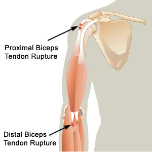
Towards the end of last year, I wrote a blog about an Achilles tendon rupture. A similar type of injury can occur in the upper arm to the biceps tendon, where this tendon is torn off the humerus bone in the upper arm. The biceps tendons attach the biceps muscles to the bone at the shoulder joint (long and short head of biceps tendon) and at the elbow joint.


Causes
Biceps tendon rupture is either caused by chronic degeneration of the tendon, or by an acute traumatic injury. Prolonged tendinitis of the biceps tendon, as a result of excessive prolonged overhead movements, such as swimming, make the tendon weak and more susceptible to tearing. In biceps tendon ruptures at the shoulder, the rupture tends to occur as a result of the tendon continually passing over the head of the humerus, causing degeneration of the tendon. This is more commonly seen in older individuals who have participated in activities involving repeated overhead movements of the arm over a long period of time. Alternatively, the biceps tendon can rupture during a traumatic event, such as heavy weightlifting.

Signs and Symptoms
An individual may hear and/or feel a snapping sensation at the site of the tear and there is usually intense pain. The following may also be present:
·         Skin discolouration and bruising in the upper arm
·         A visible abnormality in the muscle belly when the elbow is bent
·         In the case of a rupture of the long head of the biceps tendon at the shoulder, there is an obvious “Popeye” appearance, where the muscle belly bulges out (like Popeye when he flexes his biceps)
·         Ruptures at the elbow result in pain and weakness near the elbow, especially when bending the elbow and turning the palm upwards

Treatment and Management
In a complete biceps tendon rupture, surgery is required to reattach the tendon to the bone, especially in younger, more active individuals. A sling is then used to ensure the arm remains immobilized while the tendon heals. Physiotherapy is required to regain range of motion in the shoulder joint. Finally, biokinetic therapy is essential to final phase rehabilitation, to provide stretching and strengthening exercises in order to regain full function. In the less active population, surgery is not necessary, as adequate function can be regained with the help of an appropriate exercise rehabilitation programme.

References
Foundations of Athletic Training: Prevention, Assessment and Management